How Wearable EEG Could Predict Migraines: An Interview with Annemijn Oosterlee

Types of EEG Electrodes: Gel, Water, and Dry
Explaining the differences, advantages, and disadvantages of various EEG electrode types: Gel, Water, and Dry.

Multimodal Measurement: Integration of EEG & fNIRS
The integration of EEG and fNIRS combines the strengths of both techniques, capturing electrical activity and hemodynamic responses for a deeper understanding of brain function. This multimodal approach enhances research in areas like brain-computer interfaces, hyperscanning, and clinical studies, offering high temporal and spatial resolution in a non-invasive, portable setup.

Hyperscanning to Investigate Human Social Interactions
Hyperscanning is an innovative technique that allows researchers to simultaneously record and analyze the brain activity of multiple individuals during social interactions. By utilizing methods such as EEG, fMRI, and fNIRS, hyperscanning provides insights into the neural mechanisms underlying cooperative and competitive behaviors. This approach enhances our understanding of how brains communicate during shared activities, offering valuable perspectives in fields like neuroscience, psychology, and social sciences.

Unipolar vs. Bipolar EEG Measurement
In the field of electrophysiological measurements, we distinguish two types of measurement principles: the bipolar and the unipolar measurement principle. Here we will explain the difference between the principles as well as the different unipolar measurements that exist.

Event-Related Potentials in EEG
ERP is the neural response associated with a specific sensory, cognitive, or motor event (e.g. a stimulus). An ERP is often recorded using scalp electroencephalography (EEG) and looks at the average change in voltage over time starting at the onset of the stimulus over multiple trials. ERP measurements have a precise temporal resolution, which is useful in testing perception and attention.1 This information can be used to evaluate brain functioning by looking at how the brain normally processes information, as well as viewing how this processing may differ in neurological or psychiatric disorders.
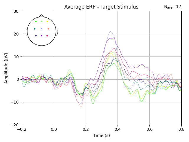
What is the P300 in Event-Related Potentials (ERPs)?
This blog describes what the P300 is and how you can detect it. It includes a practical example of how a P300 response was measured using TMSi's SAGA, including the experimental protocol, a sample dataset, and all acquisition and processing codes. At the end of this blog, you will be able to download this sample data set and run through the scripts to view the P300 yourself.

Motion Artifacts on EEG
A common problem in measurement setups where the subject is allowed to move around is the movement artifact. Movement artifacts on EEG measurements originate from two different phenomena: the movement of the cables and the movement of the electrode.
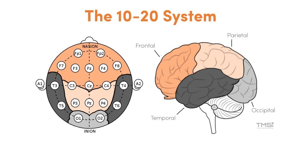
What Is the 10-20 System for EEG?
The 10-20 system is an internationally recognized method for standardizing the placement of EEG electrodes on the scalp. This system ensures consistent and replicable EEG recordings by using specific anatomical landmarks to determine electrode positions, facilitating accurate assessment of brain activity across various regions. Extensions of this system, such as the 10-10 and 10-5 systems, offer higher resolution measurements by incorporating additional electrodes between existing positions.

What Are the Different Types of Brain Waves?
This blog post explores the different types of brain waves—alpha, beta, theta, and delta—and their unique frequencies, functions, and associations with relaxation, focus, creativity, and sleep.

Mains Interference
Electrophysiological measurements often include interference from mains electricity, typically 220 V/50 Hz in Europe and 110 V/60 Hz in other regions. Even when using battery-powered amplifiers, mains interference can still affect recordings due to various pathways. Understanding these interference mechanisms is key to minimizing their impact.

Basics of Measuring Bioelectricity in humans
Bio-electricity refers to the measurable potential differences between two points on the body, providing valuable insights into the body's electrical activity. Understanding the origins of these potentials and the challenges involved in measuring them is essential for accurate interpretation and application.
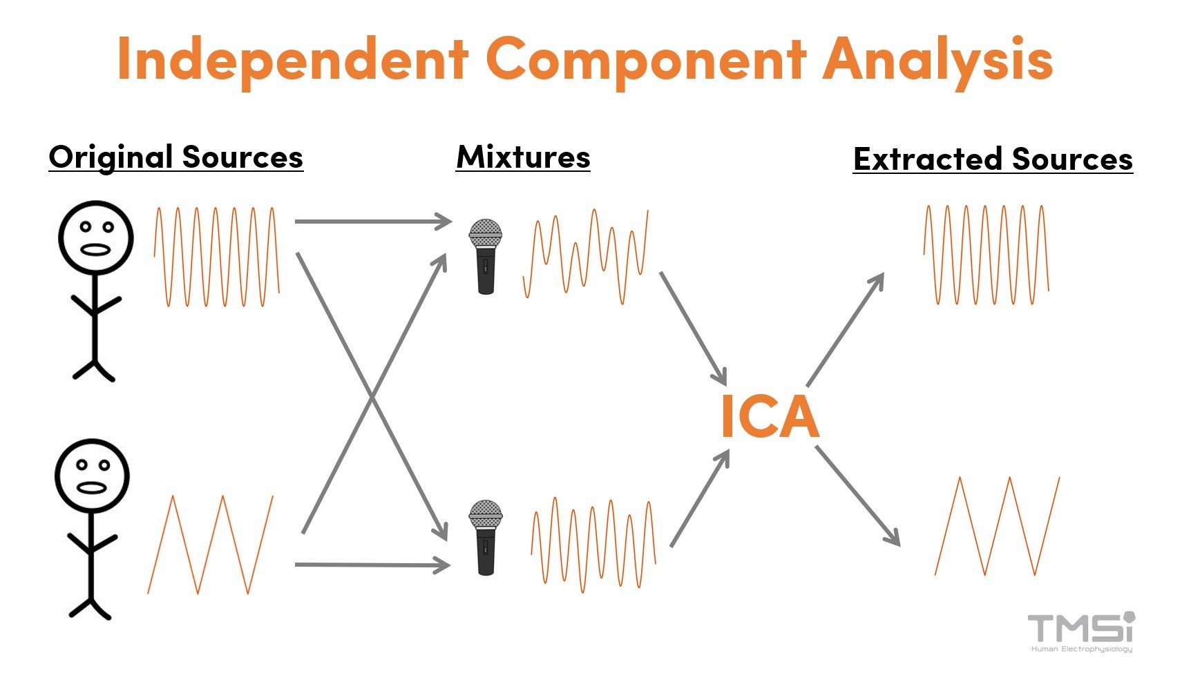
Removing Artifacts From EEG Data Using Independent Component Analysis (ICA)
Eye movements and blinks cause significant EEG artifacts, with blink artifacts being much larger in amplitude than EEG signals. Preventing eye movement artifacts is challenging, as restricting blinking or gaze can increase cognitive load. Post-processing methods like Independent Component Analysis (ICA) effectively remove these artifacts while preserving the data.

Common non-physiological (external) EEG artifacts
Learn how to recognize non-physiological (external) artifacts in the electroencephalogram (EEG) and how to prevent them.

Common physiological EEG artifacts
A guide on how to recognize physiological artifacts in the electroencephalogram (EEG) and how to manage them.

What is Neurofeedback?
Neurofeedback is a method that uses real-time brain activity to provide individuals with feedback on their brain function. Brain activity is often measured with electroencephalography (EEG). The goal of neurofeedback is to help individuals gain more control over their brain function. In this process, the user is provided with positive feedback for desirable brain activity and negative feedback for undesirable brain activity. This feedback mechanism is hypothesized to enable users to train and regulate their brain activity over time.

Valentina Barone: Predicting Outcomes in Neurological Disorders
In this user story, we would like to highlight a talented researcher: Valentina Barone. Three years ago, she started her PhD at the University of Twente in clinical neurophysiology, in collaboration with TMSi. She uses TMSi’s SAGA amplifier to record EEG signals to make a prediction of outcomes in neurological disorders. Therefore, we are happy to share her interesting research.